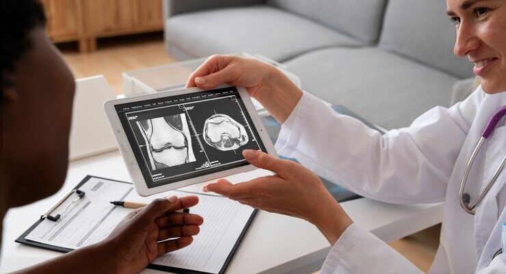
Computer Tomo urogram (CTU) and Intravenous Pyelogram (IVP) are both diagnostic methods for assessing kidney dynamics. A CT Urogram procedure retains the basic premise of a urogram but uses computed tomography to offer detailed images of the kidneys, ureters, and bladder.
On the other hand, an IVP employs contrast to make the structures outlined visible. Understanding the difference between the two is essential since they are different and yield unique information that is applicable in various clinical settings.
The Key Differences Between CT Urogram and IVP
The key differences between CT Urogram and IVP are important for healthcare professionals to consider when choosing the best option for individual patients:
- Accuracy:
CTU and IVP belong to diagnostic imaging, but they differ in accuracy. Housed at the CTU, computed tomography technology is employed in imaging the urinary tract better than ultrasound in that it has a higher accuracy coupled with the sensitivity to pick up even small lesions or stones. Again, this is a clear contrast to the IVP since that test is based upon X-ray imaging, which usually may not be as effective when identifying small pathologies.
- Invasiveness:
Research also reveals that IVP is a more invasive study than CTU, considering that it requires catheterization of the bladder with the infusion of the contrast material through the intravenous line. CTU, on the other hand, is not an invasive test that involves examination of a body and particularly depends on its pattern of defecation.
- Radiation Exposure:
IVP exposes patients to a higher dose of ionizing radiation compared to CTU. CTU uses a lower radiation dose, especially with multi-detector CT scanners and advanced imaging protocols. However, both procedures still risk radiation exposure, and steps should be taken to minimize this risk.
- Image Quality:
CTU provides high-quality images with excellent resolution, allowing for detailed visualization of the urinary tract and adjacent structures. IVP images can be of lower quality, especially in patients with severe kidney disease or those who are not able to excrete the contrast material effectively.
When to Choose a CT Urogram vs. an IVP
When deciding between a CT urogram and an IVP, there are several factors to consider:
When to prefer a CT urogram over an IVP:
- Imaging complex renal structures: CT Urogram seems more useful in imaging multiple renal arteries, horseshoe kidneys, and renal malformations.
- Detection of urinary tract stones: CT Urograms are superior in assessing UT stones, especially small, barely visible stones of less than 2mm that may not be clearly visualized on IVP.
- Evaluation of neoplastic lesions: Compared with IVU, CT Urogram provides better sensitivity in diagnosing and describing the character of neoplastic lesions in the renal parenchyma of patients, including renal cell carcinoma and metastases.
- Patients with kidney disease or renal impairment: IVP is a more significant choice for patients with kidney disease or renal impairment since this test causes contrast-induced nephropathy in patients’ conditions.
- Patients with severe allergies: However, in patients with severe iodine contrast agent allergies, a CT Urogram with non-ionic contrast materials is safer.
When to prefer an IVP over a CT urogram:
- Emergency evaluation of acute flank pain: The IVP is usually ideal in the emergency setting to evaluate acute flank pain as it offers a method of assessing the urinary tract for any obstruction or injury.
- Evaluation of vesicoureteric reflux: IVP is more suitable in the assessment of vesicoureteric reflux because it gives a clear outline of the urinary tract, and thus, the pattern of reflux is assessed.
- Low-risk patients with normal renal function: Such a choice may be justified in patients with normal kidney function and with a low probability of developing serious adverse effects linked with IVP because it causes less radiation exposure and can have fewer associated complications.
- Evaluation of renovascular disease: IVP can give more specific information about a treatment, which might include stenting or angioplasty.
- Resource constraints or limited availability of CT equipment: In cases where advanced equipment like CT scanners is hard to come by, or there is a cost constraint, IVP may be more within reach/feasible.
Are there any risks associated with CT Urogram and IVP procedures?
Risks associated with CT Urogram (Computed Tomography Urogram) and IVP (Intravenous Pyelogram) procedures:
- Allergic reactions: Patients may experience some side effects such as itching, hives, or difficulty in breathing, traceable to the contrast dye used in such processes.
- Nausea and vomiting: The contrast dye can give you stomach upset, nausea if it is not properly hydrated during the procedure.
- Kidney damage: The iodine-based contrast dye harms the kidneys, although normal and temporarily reversible kidney damage is seen in most patients, especially those with a pre-existing proven self-limiting renal impairment.
- High blood pressure: The contrast dye can occasionally raise blood pressure, and this can be unhealthy for those patients with uncontrolled hypertension.
- Increased risk of infection: It’s important to ensure everything is clean and safe when working in the patient’s private area.
What are the common findings from CT Urogram and IVP imaging studies?
Based on common radiology reports and studies, here are the common findings from CT Urogram and IVP (Intravenous Pyelogram) imaging studies:
- Stones: CT scans and IVP studies frequently show findings in the kidney area, such as blockages, movement of kidney stones, or the presence of stones.
- Hydronephrosis: Both investigations frequently note patients with distal dilatation of the collecting systems, such as the calves, pelvis, or ureter, secondary to obstruction or compression.
- Ureteral Calculi: When performing a CT Urogram and an IVP, doctors often discover small stones or obstructions in the tube leading to the bladder, particularly in the latter portion of it.
- Pyelocalyceal Dilatation: In addition, mild to moderate dilation of the kidney’s drainage system is probable on CT Urogram and IVP.
- Narrowing or Stenosis: Narrow or tight areas in the tube connecting the kidney to the bladder are often seen on certain medical imaging tests, such as CT urograms and IVP.
How should patients prepare for a CT urogram or IVP procedure?
Here’s a step-by-step guide on how patients should prepare for a CT Urogram (CTU) or Intravenous Pyelogram (IVP) procedure:
- Fasting: Patients should not eat or drink anything except medication and water for 4-6 hours before the procedure. This helps to prevent any allergic reactions or GI issues during the study.
- Avoid dairy products: Some patients may be instructed to avoid certain forms of diet, including milk products like milk, cheese, and ice cream, for about 24 hours before the procedure because of an allergy.
- Wear comfortable clothing: The patient is recommended to wear loose and light clothes as these will have to be removed during the procedure.
- Avoid using lotions or oils: Cream, oil, or powder should not be used for the skin as they interfere with the imaging modalities.
- Remove jewelry and metal objects: Necklaces and any piece of metal trinketry or glasses must be removed as they affect the quality of the picture (for instance, hearing aid, crown).
- Arrive early: The preferable time to arrived at the hospital is half an hour prior to the scheduled procedure in order to complete any forms that might have been necessary and then be comfortable.
- Inform your doctor: Inform your physician of any alterations in your health, changes in your medication regime, and allergies since they last saw you.
Conclusion
In conclusion, whenever there is a kidney problem, CT urogram and IVP assist in diagnosing and assessing the issue’s progress. Patients, practitioners, and other stakeholders must clearly distinguish the two methods concerning accuracy, invasiveness, radiation dosage, and imaging capability for patients’ better outcomes. Depending on the chosen procedure, patients with kidney problems can receive proper and adequate care from healthcare workers.


