Types of Imaging
In medicine, peering inside the human body to diagnose and treat ailments has traditionally been a complex endeavor. However, advancements in medical technology have brought forth a revolution—the art of medical imaging.
These techniques offer a noninvasive or minimally invasive window into the body, allowing healthcare professionals to visualize organs, bones, tissues, and blood flow with exceptional detail. So, let’s find out about the various types of affordable imaging used in modern medicine, explaining their principles, applications, and advantages.
X-ray: The Stalwart of Medical Imaging
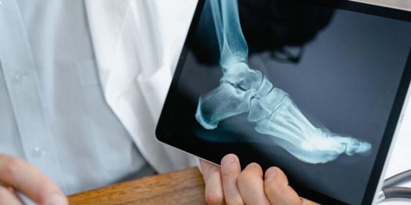
X-rays, the pioneers of medical imaging, utilize electromagnetic radiation to create internal body pictures. A regulated burst of X-rays is sent into the body, and a film or digital detector records the amount of radiation absorption by various tissues. Softer tissues, like muscles, appear gray in the picture, whereas denser tissues, like bones, absorb more X-rays and look white. X-rays are widely popular for:
Fracture diagnosis: X-rays are the gold standard for identifying broken bones due to their ability to depict bone structures clearly.
Chest X-rays: These are routinely employed to assess lung health and diagnose conditions like pneumonia, tuberculosis, and lung cancer.
Abdominal X-rays: While not as detailed as other modalities, X-rays of the abdomen can help identify blockages, inflammation, and foreign objects.
Advantages: When compared to other imaging methods, X-rays are rapid, painless, accessible, and reasonably priced.
Limitations: X-rays cannot distinguish between soft tissues with similar densities. Additionally, excessive X-ray exposure can pose health risks.
CT Scan: Unveiling a Cross-Sectional View
Computed tomography (CT) scans, often known as CAT scans, expand on the notion of X-rays. A CT scanner creates precise cross-sectional images of the body by combining several X-ray pictures acquired from various angles. A computer then reconstructs these slices to generate a three-dimensional image. CT scans provide superior detail in comparison to standard X-rays. They are useful for:
Detailed fracture assessment: CT scans can reveal intricate bone fractures and pinpoint the displacement of fragments.
Tumor detection and staging: CT scans are valuable tools for locating tumors, determining their size and spread, and monitoring treatment response.
Internal bleeding identification: CT scans can effectively detect internal bleeding in the abdomen, brain, or other areas.
Advantages: CT scans offer exceptional detail and can image various body parts. They are also relatively quick and painless.
Limitations: CT scans expose patients to ionizing radiation but at lower doses than standard X-rays. Furthermore, the contrast dye used in some CT scans may trigger allergic responses in some people.
MRI: Unveiling the Body’s Interior Through Magnetism
Magnetic resonance imaging (MRI) uses powerful magnetic fields and radio waves to produce detailed pictures of organs, soft tissues, and bones. The MRI scanner creates a magnetic field that aligns hydrogen atoms in the body’s tissues. Radio waves are then pulsed, causing these aligned atoms to absorb energy. Once the radio waves cease, the atoms release the absorbed energy, which the scanner detects and uses to construct a complete image. MRIs are particularly useful for:
Soft tissue imaging: Unlike X-rays and CT scans, MRIs excel at depicting soft tissues like muscles, ligaments, and the brain, making them ideal for diagnosing muscle tears, ligament sprains, and brain tumors.
Evaluating spinal cord injuries: MRIs provide detailed views of the spinal cord, allowing for the detection of herniated discs, spinal stenosis, and other abnormalities.
Cancer diagnosis: MRIs can aid in cancer diagnosis by identifying tumors and assessing their spread to surrounding tissues.
Advantages: MRIs do not involve ionizing radiation, making them a safer option for certain patients. They also offer exceptional soft tissue detail.
Limitations: MRIs can be claustrophobic due to the enclosed nature of the scanner. Additionally, they are not suitable for patients with certain medical implants or claustrophobia.
Ultrasound: Harnessing Sound Waves for Real-Time Imaging
Ultrasound imaging uses high-frequency sound waves to provide real-time pictures of internal tissues and organs. A transducer, similar to a microphone, is placed on the body’s surface. The transducer emits sound waves that travel into the body and bounce back upon encountering different tissues. The machine analyzes the sound waves to generate an image on the screen. Ultrasound is particularly valuable for:
Pregnancy monitoring: Ultrasound is the mainstay of prenatal care, allowing for visualization of the developing fetus, assessment of fetal growth, and detection of any abnormalities.
Abdominal imaging: Ultrasound effectively examines the liver, gallbladder, kidneys, and other abdominal organs to identify gallstones, cysts, and other issues.
Musculoskeletal imaging: Ultrasound can aid evaluation of muscles, tendons, and joints for tears, strains, and fluid buildup.
Advantages: Ultrasound is painless, radiation-free, and relatively inexpensive. It also offers real-time imaging, allowing for dynamic visualization of structures.
Limitations: Ultrasound images can be incomplete due to overlying bones or air pockets within the body. Additionally, the quality of the image depends on the operator’s skill.
Nuclear Medicine Imaging: Unveiling Function with Radiotracers
Nuclear medicine imaging techniques utilize small amounts of radioactive materials called radiotracers. These radiotracers are injected into the bloodstream or ingested and accumulate in specific organs or tissues based on their function. Special cameras then detect the emitted radiation, creating images that reveal the function and activity of various organs. Nuclear medicine imaging aids the following:
Bone scans: These scans help detect bone fractures, infections, and tumors by revealing areas of increased or decreased bone activity.
Thyroid scans: Nuclear medicine scans can assess thyroid function and identify abnormalities like nodules or hyperthyroidism.
Myocardial perfusion imaging (MPI): MPI scans evaluate blood flow to the heart muscle, aiding in the diagnosis of coronary artery disease.
Advantages: Nuclear medicine imaging provides valuable insights into organ function, which complements anatomical information from other imaging techniques.
Limitations: Nuclear medicine procedures involve exposure to low levels of radiation. Additionally, some radiotracers can cause allergic reactions in certain individuals.
Bottom Line: Choosing the Right Imaging Technique
In conclusion, the selection of the most appropriate imaging technique for a particular medical condition depends on several factors. These include the nature of the problem, the area of the body under examination, the patient’s medical history, and any potential risks associated with the imaging modality.
X-rays remain crucial for initial evaluations due to their speed and accessibility. CT scans offer superior detail for complex fractures and internal bleeding. MRIs excel at depicting soft tissues and are invaluable for neurological conditions. Ultrasounds provide real-time imaging, making them ideal for pregnancy monitoring and certain musculoskeletal evaluations.
AQ Imaginge sheds light on organ function and is crucial for assessing disorders like thyroid dysfunction or coronary artery disease. As a result, by knowing the strengths and limits of each imaging modality, healthcare providers may make more educated judgments to guarantee the best possible patient care.


